


From this article, you can learn about the characteristics of varicose veins in women in the pelvis - a deformity of the veins in the pelvic region with impaired blood flow to the internal and external genitals.
general information
Pelvic varicose veins are also referred to in the literature as "pelvic congestive syndrome, " "female varicocele, " and "chronic pelvic pain syndrome. "Pathology of pelvic veins is most commonly diagnosed in the reproductive period in patients aged 25-45 years.
In the vast majority of cases (80%), varicose transformation affects the veins of the ovaries and is extremely rare (1%) in the veins of the broad band of the uterus. According to the modern medical approach, the treatment of VVMT should be performed not so much from a gynecological point of view, but primarily from a phlebological point of view.
Causes of pathology
Under the varicose veins of the pelvic organs in women, doctors understand the changes in the structure of the blood vessel walls that are characteristic of other types of disease - weakening and then stretching and the formation of "pockets" in which the blood stagnates. Extremely rare cases involving only the vessels of the pelvic organs. Approximately 80% of patients with this form show signs of varicose veins in the inguinal veins of the lower extremities.
The incidence of pelvic varicose veins is most pronounced in women. This is due to anatomical and physiological features that suggest weakening of the venous walls:
- hormonal fluctuations, including fluctuations in the menstrual cycle and pregnancy;
- increased pressure in the pelvis, which is characteristic of pregnancy;
- periods of filling the veins with more active blood, including cyclic menstrual periods, during pregnancy and during sex.
All of these phenomena fall into the category of factors that cause varicose veins. And they are found exclusively in women. Most patients experience pelvic varicose veins during pregnancy because the provocative factors stratify simultaneously. According to statistics, varicose veins in the small pelvis are 7 times less common in men than in the better sex. They have several provocative factors:
- hypodynamics - long-term maintenance of low physical activity;
- increased physical activity, especially pulling weights;
- obesity;
- lack of sufficient fiber in the diet;
- inflammatory processes in the organs of the urogenital system;
- sexual dysfunction or deliberate denial of sexual intercourse.
Genetic predisposition can also lead to pathology of the plexuses inside the pelvis. According to statistics, varicose veins of the perineum and pelvic organs are most commonly diagnosed in women whose relatives have suffered from the disease. The first changes in them can be observed during adolescence and puberty.
In women with pelvic vascular involvement, the highest risk of developing groin varicose is observed in patients with venous pathology in other parts of the body. In this case, we are talking about the innate weakness of the veins.
Ethiopathogenesis
Proctologists believe that the following main causes always contribute to the occurrence of VVP: valvular insufficiency, venous occlusion, and hormonal changes.
Pelvic venous congestion syndrome can be caused by a congenital lack or insufficiency of venous valves, which has been revealed in anatomical studies in the last century, and this is confirmed by today's data.
Varicose veins were also found to be of genetic origin in 50% of patients. FOXC2 was one of the first identified genes to play a key role in the development of VVP. Currently, the association between disease development and gene mutations (TIE2, NOTCH3), thrombomodulin level, and type 2 transforming growth factor β has been determined. These factors contribute to a change in the structure of the valve itself or the venous wall - all of which leads to a failure of the valve structure; enlargement of the vein, which causes a change in the function of the valve; progressive reflux and ultimately varicose veins.
Connective tissue dysplasia can play an important role in the development of the disease, the morphological basis of which is a decrease in the content of different types of collagen or a violation of the ratio between them, which leads to a decrease in the strength of the veins. .
The incidence of VVP is directly proportional to the degree of hormonal changes that occur especially during pregnancy. In pregnant women, the capacity of the pelvic veins increases by 60% due to the mechanical compression of the pelvic vessels by the pregnant uterus and the vasodilatory effects of progesterone. This venous dilation persists for one month after delivery and can cause venous valve failure. In addition, the weight of the uterus increases during pregnancy, and changes in position occur, causing the ovaries to elongate and then become venous.
Risk factors also include endometriosis and other inflammatory diseases of the female reproductive system, estrogen therapy, unfavorable working conditions for pregnant women, which include heavy physical work and prolonged compulsion (sitting or standing) during the working day.
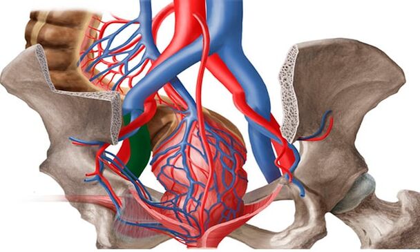
The anatomical features of the outflow from the pelvic veins also contribute to the development of the pelvic varicose vein. The diameter of the ovarian veins is usually 3-4 mm. The long and thin ovarian vein on the left flows into the renal vein on the left and the inferior vena cava on the right. Normally, the left renal vein is located in front of the aorta and behind the upper mesenteric artery. The physiological angle between the aorta and the upper mesenteric artery is about 90 °.
This normal anatomical position prevents the left renal vein from compressing. On average, the angle between the aorta and the upper mesenteric artery is 51 ± 25 ° in adults, 45, 8 ± 18, 2 ° in boys and 45, 3 ± 21, 6 ° in girls. When the angle decreases from 39, 3 ± 4, 3 ° to 14, 5 °, aorto-mesenteric compression or nut-breaking syndrome occurs. This is the so-called anterior, or true nut-breaking syndrome, which has the greatest clinical significance. Posterior necrosis syndrome is rare in patients with a retroaortic or annular arrangement of the distal left renal vein. Clogging of the proximal venous bed causes an increase in pressure in the renal vein, leading to the development of renoovarian reflux in the left ovarian vein with the development of chronic pelvic venous insufficiency.
May-Turner syndrome - compression of the left common iliac vein by the right common iliac artery - is also an etiological factor in pelvic varicose veins. It occurs in up to 3% of cases and is more common in women. Currently, due to the introduction of radiation and endovascular imaging methods, this pathology is increasingly being detected.
Classification
Varicose veins can be divided into the following forms:
- The primary type of varicose veins is an increase in the blood vessels in the pelvis. This is due to two types of key failure: acquired or congenital.
- The secondary form of pelvic venous thickening is diagnosed only in the presence of gynecological diseases (endometriosis, neoplasms, polycystic).
Pelvic varicose veins develop gradually. There are several main stages in the development of the disease in medical practice. They differ depending on the presence of complications and the spread of the disease:
- First degree. Changes in the structure of the venous valves in the ovaries can be due to hereditary or acquired causes. The disease is characterized by an increase in the diameter of the veins to 5 mm. The outer parts of the left ovary have a pronounced extent.
- Secondary. This degree is characterized by the spread of pathology and damage to the left ovary. The veins in the uterus and right ovary can also dilate. The expansion diameter reaches 10 mm.
- Third degree. The diameter of the veins increases by up to 1 cm. Varicose veins can be observed in both the right and left ovaries. This stage is due to pathological phenomena of a gynecological nature.
It is also possible to classify the disease according to the primary cause of its development. There is a primary grade in which dilation is caused by a malfunction of the venous valves and a secondary grade that is the result of chronic female disease, inflammatory processes, or complications of an oncological nature. The extent of the disease may vary depending on the anatomical feature that indicates the location of the vascular disorder:
- Plenty within the caste.
- Vulva and perineal.
- Combined forms.
Symptoms and clinical manifestations
In women, pelvic varicose veins are accompanied by severe but non-specific symptoms. Manifestations of this disease are often considered signs of gynecological disorders. The main clinical symptoms of lumbar varicose veins in women involving pelvic vessels are:

- No menstrual pain in the lower abdomen. Their intensity depends on the stage of venous damage and the extent of the process. Grade I varicose veins are characterized by intermittent, mild pain in the lower back. In the later stages it is felt in the abdomen, perineum and lower back, long and intense.
- Abundant mucous discharge. The so-called leucorrhoea does not have an unpleasant odor, it does not change color, which would indicate an infection. In the second phase of the cycle, the amount of discharge increases.
- Increased symptoms of premenstrual syndrome and dysmenorrhea. Women’s pain increases even before menstruation begins, all the way to walking difficulties. During menstrual bleeding, it can become unbearable, spreading to the entire pelvic region, the barrier, the lower back, and even the thighs.
- Another characteristic sign of women’s groin varicose veins is the discomfort that occurs during sexual intercourse. It is felt in the pubic body and vagina and is characterized as dull pain. It can be observed at the end of intercourse. In addition, the disease is accompanied by increased anxiety, irritability and mood swings.
- As in the case of pelvic varicose veins in men, the interest in sex in the female part of patients with such a diagnosis gradually disappears. The cause of the dysfunction is a permanent discomfort and a decrease in the production of sex hormones. In some cases, infertility may occur.
Instrumental diagnostics
Varicose veins are diagnosed and treated by a phlebologist and vascular surgeon. Currently, due to new technologies, the number of VVP detection cases has increased. Patients with CPP are examined in several stages.
- The first stage is a routine examination by a gynecologist: anamnesis, manual examination, ultrasound examination of the pelvic organs (to exclude other pathologies). Based on the results, a proctologist, urologist, neurologist, and other related professionals will prescribe an examination.
- If the diagnosis is unclear but VVPT is suspected, ultrasound angioscanning of the pelvic veins (USAS) is performed in the second stage. This is a non-invasive, highly informative screening method used in all women with suspected VVPT. If previously it was thought that it was enough to examine only the pelvic organs (venous examination was considered difficult to access and optional), then ultrasound examination of the pelvic veins is now a mandatory examination procedure. By measuring the diameters and the speed of blood flow in the veins, the method can be used to determine the formation of the pelvic varicose vein and to find out in advance what is the leading pathogenetic mechanism - venous failure. ovarian veins or venous occlusion. This method is also used to dynamically evaluate the conservative and surgical management of VVPT.
- The research is conducted transvaginally and transomdominally. Parametric veins, lumbar plexuses, and uterine veins are visible transvaginally. According to various authors, the diameter of the vessels of said localization is 2, 0-5, 0 mm (average 3, 9 ± 0, 5 mm), i. e. not more than 5 mm and the mean diameter of the arched veins is 1, 1 ± 0, 4 mm. Veins larger than 5 mm in diameter are considered dilated. Examination of the inferior vena cava, venous hips, left renal vein, and ovarian veins is performed transientinally to exclude thrombotic masses and extravascular compression. The left renal vein is 6-10 mm long and has an average width of 4-5 mm. Normally, the left renal vein at the point where it passes through the aorta flattens out slightly, but there is a 2-2. 5-fold decrease in its transverse diameter without a significant acceleration of blood flow, which ensures normal outflow without increasing the pressure in the prethenotic. zone. In the case of narrowing of the vein underlying the pathological compression, its diameter decreases significantly - 3. 5 to 4 times, and blood flow accelerates - above 100 cm / s. The sensitivity and specificity of this method are 78% and 100%, respectively.
- Examination of the ovarian veins is a mandatory examination of the pelvic veins. They are located along the anterior abdominal wall, along the straight abdominal muscle, slightly laterally relative to the hip veins and arteries. In the USAS, a sign of ovarian venous insufficiency is the presence of retrograde blood flow greater than 5 mm in diameter. Ultrasound examinations of the veins of the lower extremities, the perineum, the vulva, the inner thigh, and the gluteal region should be performed for a complete examination, prevention of relapses, and proper treatment tactics.
- Advances in medical technology have led to the use of new diagnostic methods. In the third stage, after ultrasound verification of the diagnosis, radiation diagnostic methods are used to confirm it.
- Pelvic phlebography with selective bilateral radiopaque ovarycography is one of the most invasive diagnostic methods performed in a hospital setting. This method has long been considered the diagnostic "gold standard" for evaluating dilatation and detecting pelvic venous insufficiency. The essence of the method is that, under the control of an X-ray machine, a contrast agent is delivered through the catheter inserted into one of the main veins (jugular, brachial or femoral) into the hip, kidney and ovarian veins. This makes it possible to identify anatomical variations in the structure of the ovarian veins and to determine the diameter of the gonads and pelvic veins.
- The retrograde contrast of the genital veins at the height of the Valsalva test serves as a pathognomic angiographic sign of valvular failure by visualizing sharp dilation and curvature, respectively. This is the most accurate method for detecting thrombophlebitic lesions of May-Turner syndrome, iliac, and vena cava.
- When the left renal vein is compressed, perirenal venous collaterals are defined with retrograde blood into the gonadal veins, contrast stagnation in the renal vein. The method measures the pressure gradient between the left kidney and the inferior vena cava. Usually 1 mmHg. Art. ; gradient 2 mmHg. Art. , May suggest slight compression; With a gradient greater than 3 mmHg. Art. high blood pressure in the left renal vein can be diagnosed with aortic mesenteric compression syndrome and the gradient5 mmHg. Art. it is considered a hemodynamically significant narrowing of the left renal vein. Determining the pressure gradient is an important element of diagnostics, as depending on its values, significantly different surgical interventions are planned in the pelvic veins, which is very important in modern conditions. Currently, this assay (with a normal pressure gradient) can be used for therapeutic purposes to embolize ovarian veins.
- The next irradiation method is emission computed tomography of pelvic veins with labeled erythrocytes in vitro. It is characterized by the deposition of labeled erythrocytes in the pelvic veins and the display of gonadal veins, allowing the identification of varicose veins of the pelvis and dilated ovarian veins at different positions, the degree of venous congestion in the pelvis, and the return of blood to the pelvic veins. Normally, the ovarian veins are not contrasting, and accumulation of radiopharmaceuticals in the venous plexuses is not observed. For an objective assessment of the degree of venous stagnation of the pelvis, the venous stagnation coefficient of the pelvis is calculated. But this method also has disadvantages: invasiveness, relatively low spatial resolution, the impossibility of accurately determining the diameter of the veins, which is why it is not used so often in clinics at present.
- Video laparoscopic examination is a valuable tool in the evaluation of undiagnosed. In combination with other methods, it can help determine the causes of pain and prescribe appropriate treatment. In the ovarian region, the veins along the round and wide strips of the uterus with the veins of the pelvis can be displayed in the form of cyanotic, dilated vessels, tapered, and tense walls. The use of this method is significantly limited by the presence of retroperitoneal adipose tissue, the possibility of assessing varicose veins only in a limited area, and the impossibility of detecting venous reflux. Currently, the use of this method is diagnosed when multifocal pain is suspected. Laparoscopy allows the visualization of the causes of CPP, such as foci of endometriosis or adhesions, in 66% of cases.
Characteristics of therapy
To fully treat varicose veins in the small pelvis, the woman should follow all of the doctor’s recommendations and change her lifestyle. Above all, weights should be paid attention to if they are too high, they should be reduced if the patient is living an excessively sedentary lifestyle, exercising, walking more often, and so on.
People with varicose veins are strongly advised to change their diet as little as possible fast food (fried, smoked, sweet in large quantities, salty, etc. ), alcohol, caffeine. It is better to give preference to vegetables and fruits, dairy products, cereals.
In addition, to prevent the progression of the disease and for medical purposes, doctors are required to wear compression underwear for patients with varicose veins.
Medicines
ERCT therapy involves a number of important points:
- get rid of the reverse flow of venous blood;
- relieving the symptoms of the disease;
- stabilization of vascular tone;
- improved blood circulation in the tissues.
Preparations for varicose veins should be taken in courses. Other painkillers should only be taken during a painful attack. To be effective in therapy, your doctor will often prescribe the following medications:
- phleboprotectors;
- enzyme preparations;
- drugs that relieve inflammatory processes in varicose veins;
- tablets to improve blood circulation.
Operational treatment
It is worth recognizing that conservative treatment methods give really visible results primarily in the early stages of varicose veins. However, the problem can basically be solved and the disease can be completely eradicated only with surgery. There are many variants of surgical treatment of varicose veins in modern medicine, consider the most common and most effective types of surgery:
- embolization of veins in the ovary;
- sclerotherapy;
- plastic for uterine bands;
- removal of enlarged veins by laparoscopy;
- clamping of veins in the pelvis with special medical clips (cutting);
- crossectomy - ligation of veins (which, in addition to the pelvic organs, also affects the vessels of the lower extremities).
Symptomatic treatment of pelvic varicose veins during pregnancy is only possible. We recommend wearing compression stockings, phlebotonics on the recommendation of a vascular surgeon. Phlebosclerosis of the perineum varicose veins can be performed in the II-III trimester. If there is a high risk of bleeding during spontaneous delivery due to varicose veins, a decision should be made for surgical delivery.
Physiotherapy
The system of physical activity for the treatment of varicose veins in women consists of exercises:
- "Bicycle". We lie on our backs, lay our hands behind our heads, or place them along our body. Lifting our legs, we make circular movements with them, as if we were pedaling on a bicycle.
- "Birch". We sit face up on any hard, comfortable surface. Raise your legs and gently launch them behind your head. Support the lumbar region with your hands and place your elbows on the floor, slowly straighten your legs, lift your body.
- "Scissors". The starting position is in the back. Raise the closed legs slightly above floor level. The lower limbs are laid out to the side, put back on and repeated.
Possible complications
Why are pelvic varicose veins dangerous? The following consequences of the disease are often recorded:
- inflammation of the uterus, its appendages;
- uterine bleeding;
- disorders in the work of the bladder;
- development of venous thrombosis (small percentage).
Prophylaxis
In order to prevent the pelvic varicose vein from disappearing as soon as possible and to prevent the pathology of the pelvic organs from recurring in the future, it is advisable to follow simple prevention rules:
- perform gymnastic exercises daily;
- prevents constipation;
- follow a diet in which plant fiber must be present;
- do not stay in one position for long;
- take a contrast shower from the perineum;
- to prevent varicose veins from appearing, it is worth wearing exceptionally comfortable shoes and clothing.
Preventive measures to reduce the development and progression of pelvic varicose veins are mainly limited to the normalization of lifestyle.